Basal Cell Carcinoma
Where Does Basal Cell Carcinoma Come From?
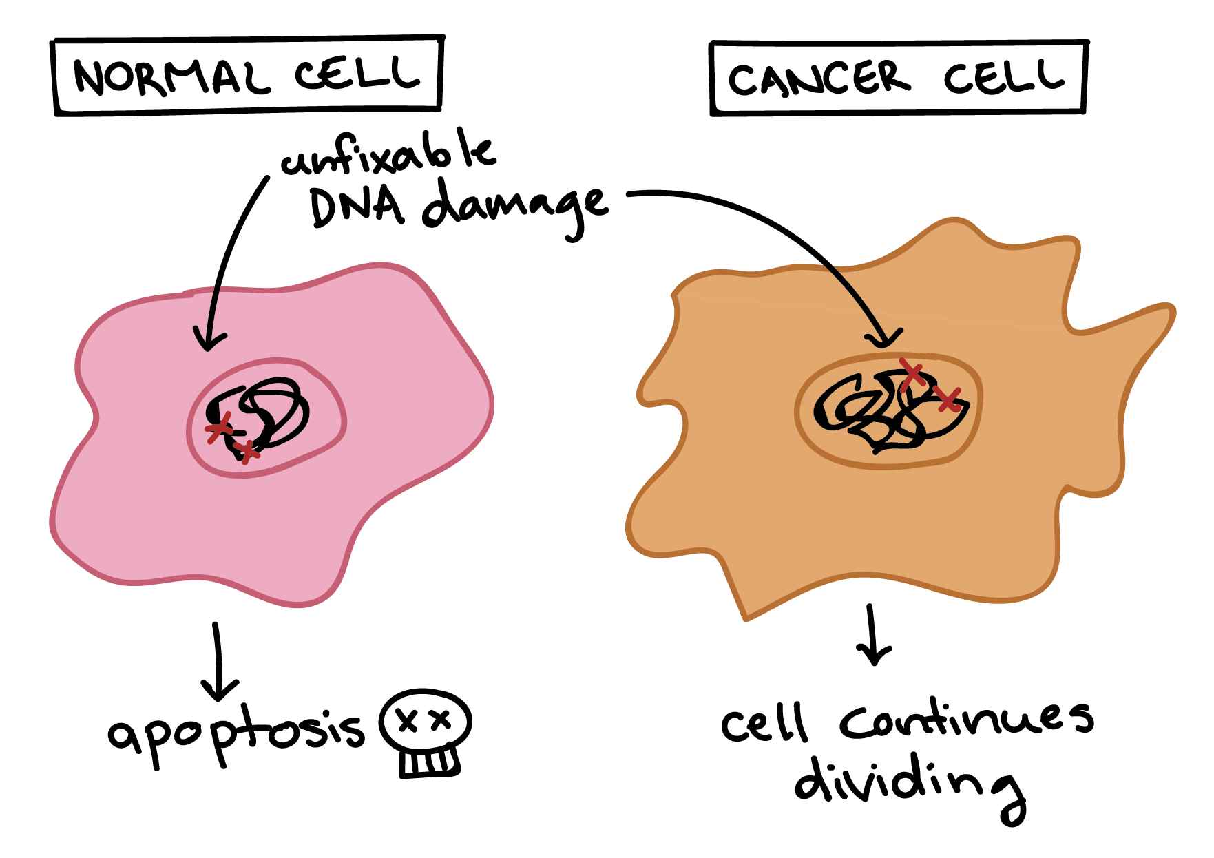
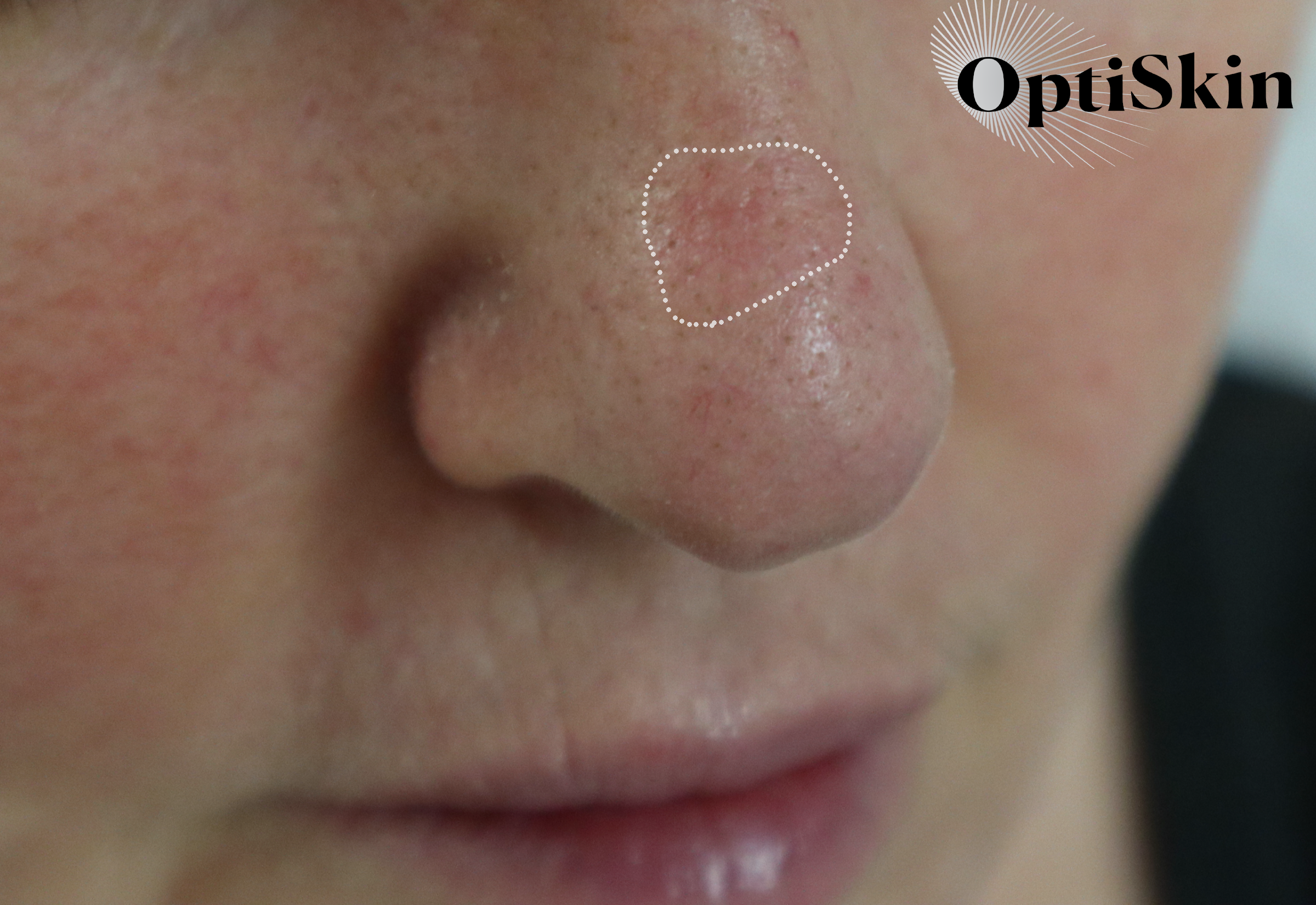
The most common type of skin cancer in the United States, basal cell carcinoma (commonly referred to as BCC) is caused by a mutation in the DNA of skin cells in the deepest (basal) layer of the epidermis. BCC is typically the result of extended UV exposure and is thus found in areas regularly exposed to the sun, such as the ears, eyes, nose, and scalp.
Who Is Most at Risk of Developing BCC?
Although people with all skin types can develop basal cell carcinomas, UV exposure is the number one cause of BCC, and if you are fair-skinned, have a family history of skin cancer, have ever had a sunburn that blistered, or you have a weakened immune system, you are considered high risk for BCC.
Is BCC Life-Threatening?
No, BCC is usually not life-threatening. But removal of an advanced BCC tumor can be disfiguring. So, please, don’t skip those annual skin checks!
What Does BCC Look Like?
Not all BCCs look the same. These are the four most common types.
Nodular BCC is the most common, and it usually looks like a pink pearly papule. Patients often describe a nodular BCC as ‘a new pimple that started to bleed and will not heal.’ This type of lesion responds very well to treatment with non-ablative lasers.
Superficial BCC is the second most common type of BCC, and it usually looks like a pink or reddish patch. This type of lesion shows up vividly when viewed with a dermatoscope (a handheld microscope), and boasts a leaf-like structure. This type of BCC responds best to topical creams such as Aldara, as well as treatment with a non-ablative laser.
Infiltrative BCC can penetrate quite deeply into the dermis, making this type of BCC more apt to permanently damage the skin and scar. Viewed through advanced imaging devices, infiltrative BCC often appears as poorly defined red or ulcerated plaque. Infiltrative BCC is important to catch and manage early because these lesions do grow quickly and and can be difficult to remove surgically. The good news is, when caught early, they respond well to treatment.
Pigmented BCC contain melanin and may appear as a dark spot, especially in skin of color. Because of their dark color, pigmented BCC are usually fairly easy to spot using a dermatoscope, and recent research done by Dr. Markowitz shows that they do respond well to non-ablative laser treatment.
If you suspect you may have a BCC spot, be proactive and visit OptiSkin so Dr. Markowitz can take a look. Remember, early intervention is key to minimizing the invasiveness (and scarring) of any skin cancer treatment.
How We Diagnose BCC at OptiSkin
Dr. Markowitz does not excise suspicious spots with a scalpel and send them off to a lab to be analyzed. Thanks to her state-of-the-art imaging devices, Dr. Markowitz is able to diagnose most skin cancer tumors noninvasively. At every skin exam, Dr. Markowitz first utilizes a dermatoscope to magnify the skin and study every inch in detail. If she does see an area that merits closer investigation, Dr. Markowitz will then use her confocal microscope to capture a more detailed image. This image enables her to identify mutations in the skin cells and diagnose skin cancer likes BCC without ever having to cut the skin. Using the imaging devices also enables Dr. Markowitz to identify BCC earlier—well before it presents on the skin as sore or bump.
How We Treat BCC at OptiSkin
Dr. Markowitz creates a customized treatment regimen for each patient. She will typically first analyze the size and depth of the skin cancer tumor using optical coherence tomography (a noninvasive laser), then she designs a protocol of non-ablative lasers and topicals to treat the area. For most patients, there is little to no downtime while treating BCC, and many tumors are resolved in just one to two treatments.
Does BCC Ever Go Away or Shrink?
No. Your system may keep a BCC tumor contained for years, but, eventually, it will erupt and invade surrounding areas. This is why routine skin checks are so important.
Call OptiSkin Today
If you are concerned about a spot on your skin—or have any of the risk factors outlined above—alleviate your worry by calling today to schedule an appointment with Dr. Markowitz. BCC is highly treatable, but the goal is to catch it early and minimize the invasiveness of treatment.
Not all BCCs look the same—and not all are visible to the naked eye. That is why Dr. Markowitz utlizizes advanced imaging devices to study the skin at a microscopic level, enabling her to differentiate between superficial, nodular and infiltrative BCC. Accurately identifying the type of BCC present is important because it informs the course of treatment. Copyright OptiSkin 2021. Artwork by Alisa Girard @artsymed
Harmful UV rays from the sun can cause irreversible damage to a cells DNA, leading to either cell death or the development of cancer.
Artwork by Khan Academy.
A nodular BCC with ulceration, these often appear as a pink pearly bump and may bleed or ulcerate.
Superficial Spreading Basal Cell Carcinoma, under magnification and illumination with a dermatoscope.
Focal Sclerosing Basal Cell Carcinoma may appear as a red patch with some scarring or discoloration, but these tumors are highly invasive and can destroy large parts of the face before they are fully removed.
An example of pigmented basal cell carcinoma as viewed with the naked eye and on dermoscopy.
©Optiskin 2021


